Assay Readouts
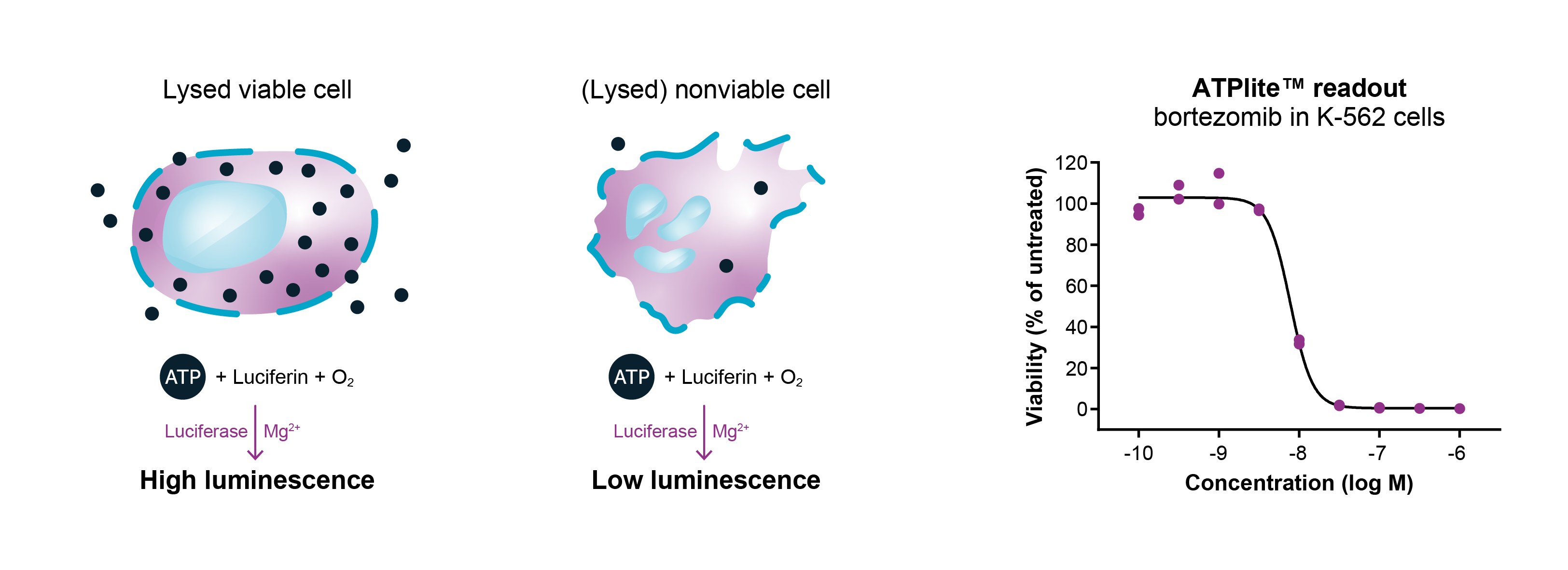
ATPlite™
We use ATPlite™ (Revvity) as the standard readout for cell viability assays. This high-throughput luminescence assay is based on the determination of ATP content using firefly luciferase. As all living cells contain ATP, this molecule can serve as an indirect marker of cell number. After lysis of cultured cells, addition of luciferase and luciferin produces light, upon reaction with ATP. This emitted light is proportional to the ATP concentration, thereby allowing quantitative determination of the effect of compound on cell viability. By comparing the signal after incubation of cells with a compound for a number of days, its effect on cell proliferation or cell toxicity can be determined. We use the ATPlite™ assay to investigate the effect of both small molecules and biologics on (cancer) cell viability.
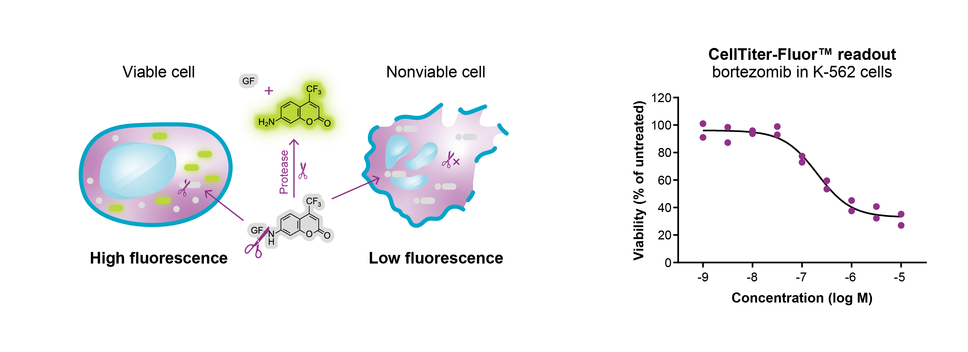
CellTiter-Fluor™
In addition to our standard ATP readout, we offer other readouts for cell viability, including CellTiter-Fluor™ (Promega). This high-throughput fluorescence assay is based on measurement of constitutive protease activity that is present in viable cells. This non-lytic method makes use of a fluorogenic peptide substrate that can cross the membrane of intact cells. Once inside the cell, the peptide substrate is cleaved by protease activity, resulting in a fluorescent signal. This signal is proportional to the number of viable cells. Low fluorescence will be observed in case of dying or dead cells as the live-cell protease will have leaked into the culture medium and has become inactive.
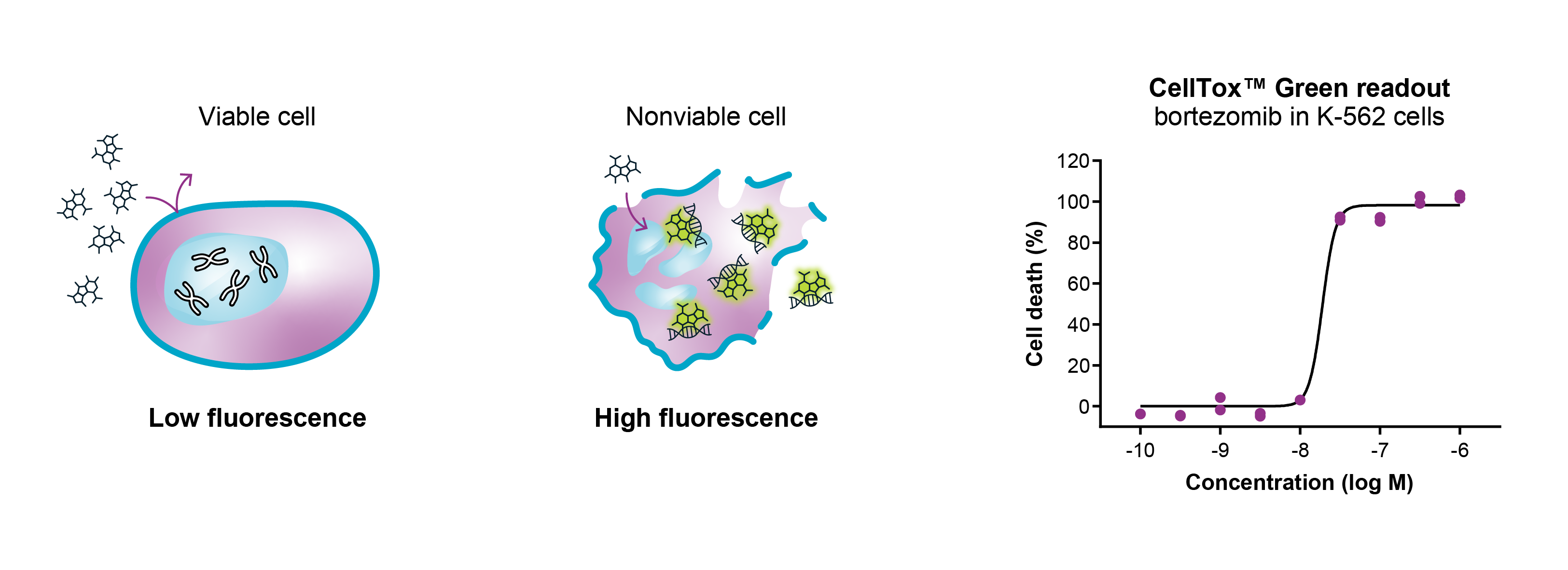
CellTox™ Green
While the ATPlite™ and CellTiter Fluor™ assays are used for measurement of cell viability, the CellTox™ Green assay (Promega) allows detection of changes in the membrane integrity upon cell death. This non-lytic, high-throughput assay utilizes a cyanine dye to evaluate cytotoxicity in cultured cells after treatment with small-molecule compounds or biologics. In case of viable cells, the cyanine dye cannot cross the impermeable cell membrane, resulting in a low fluorescence signal. When the cell membrane integrity is lost in dying or dead cells, the dye can enter the cell and bind to the DNA. Upon formation of the cyanine-DNA complex, the fluorescence signal increases.

Information Needed?
For more information on Oncolines® proliferation assays and bioinformatics analysis please contact us.